* Dermoid Sinus
* Demodectoc Mange
(Localized and Generalized)
(Localized and Generalized)
RIDGEBACK HEALTH
WHAT IS COCCIDIA?
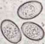
Coccidia are small protozoans(one-celled organisms) that multiply in the intestinal tracts of dogs, most commonly in puppies less than six months of age, in adult animals whose immune system is suppressed or in animals who are stressed in other ways (change in ownership, other disease present, stress, etc.). In dogs, most coccidia are of the genus called ISOSPORA. Isospora canis and I. Ohioans are the species most often encountered in dogs. As a puppy ages it tends to develop a natural immunity to the effects of coccidia. As an adult it may carry coccidia in its intestines, shed the cyst in the feces, but experience no ill effects.
HOW ARE THE COCCIDIA TRANSMITTED?
Coccidia is carried from yard to yard by infected birds, droppings, feathers and flies. If one puppy has coccidia, usually all the puppies will, by fecal matter on the rectum and from stepping in another puppies fecal matter. A Puppy is not born with coccidia organisms in its intestines. However, once born, the puppy is frequently exposed to its mothers feces and if the mother is shedding the infective cysts in her feces, then the young puppies will likely ingest them and coccidia will develop within their intestines. Since young puppies usually those less than six months of age have no immunity to coccidia, the organisms reproduce in great numbers and parasitize the young puppies intestines. Oftentimes this has severe effects.
WHAT ARE THE SYMPTOMS OF COCCIDIA?
The primary sign of coccidia is diarrhea. The diarrhea usually has a foul smelling odor. The diarrhea may be mild to severe depending on the level of infection. Blood and mucous may also be present, especially in advanced cases. Severely affected animals may also vomit, lose their appetite, become dehydrated, and in some cases, if not treated, die from coccidia. Most infected puppies usually are in the four to twelve age group. The possibility of coccidia should always be considered when a loose stool or diarrhea is encountered in this age group. A microscopic fecal exam by a veterinarian will detect the cysts confirming a diagnosis.
WHAT ARE THE RISKS?
Although most cases of coccidia are very mild, it is not uncommon to see severe, bloody diarrhea resulting in dehydration and even death. Coccidia is most common to animals who are ill or infected with other parasites, bacteria or viruses. Coccidia is very contagious from the age group of four to twelve week age group in puppies. It should also be mentioned that STRESSplays a big role in the development of coccidia. It is not uncommon for a seemingly healthy puppy to arrive at its new home and develop diarrhea several days later leading to a diagnosis of coccidia. Coccidia can surface during the stressful period of the puppy adjusting to a new home. Fortunately coccidia is treatable. Drugs such as sulfadimethoxine (Albon), trimethoprimsuladiazine (Tribrissen)and amprolium (Corid) have all been effective in the treatment and prevention of coddidia. and amprolium (Corid) have all been effective in the treatment and prevention of coddidia. Because these drugs DO NOT kill the organisms, but rather inhibit their reproduction capabilities, elimination of coccidia from the intestine is not rapid. By stopping the ability of the protozoa to reproduce, time allowed for the puppys own immunity to develop and remove the organisms. Drug treatments of five or more days are usually required. Puppies are no longer contagious after treatment for two days.
HOW IS COCCIDIA PREVENTED OR CONTROLLED?
Coccidia use to be thought of as a parasite that only came from unclean kennels. Now we know that it can be carried in by people, dogs and fleas as well. Adults dont always have symptoms and can be carriers of Coccidia. Because coccidia is spread by the feces of carrier animals, it is very important to practice strict sanitation. Clean puppy cages often and bath all pups and adults daily until gone. All fecal material should be removed. Housing needs to be such that the food and water cannot become contaminated with feces. Clean water should be provided at all times. Most disinfectants do not work well against coccidia. Incineration of the feces, steam cleaning, immersion in boiling water or a 10% ammonia solution are the best methods to kill coccidia. Coccidia can withstand freezing. Cockroaches and flies can mechanically carry coccidia from one place to another. Mice and other animals can ingest the coccidia and when killed and eaten by a cat, dog, for instance, can infect the cat or dog. Therefore, insect and rodent control are very important in preventing coccidia. The coccidia species of dogs and cats do not infect humans.
* Coccidia
|
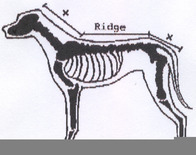
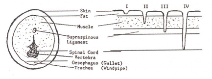
DERMOID SINUS IN THE RHODESIAN RIDGEBACK (A Review by a Veterinarian) Taken from South African Rhodesian Ridgeback Club, Ridgeback ReviewTHE FORMATION AND SIGNIFICANCE OF DERMOID SINUSThe Rhodesian Ridgeback is a modern breed of dog that originated in the late nineteenth century, by the crossing of indigenous Hottentot dogs with various European breeds introduced into the Cape by the early settlers.The breed standard was established with the formation of the Rhodesian Ridgeback Club of Bulawayo in 1922. The main characteristics of the breed is, as its name implies-a ridged back, which is formed in the haircoat along the top midline of the dog's back. The ridge is formed by hair, which grows in the opposite direction to the hair of the surrounding coat.Breeders of Ridgebacks are aware of a well-known defect which occurs in the breed, the Ridgeback "Cyst" or as it is more correctly named in the scientific terminology, the Dermoid Sinus. (Dermoid-arising from the skin, Sinus-a cavity or channel).Dermoid Sinuses are narrow tube-like structures, which are derived from a skin defect. They penetrate from the skin surface to varying depths downward into the muscles and towards the spinal cord. They are situated in the midline of the neck and croup, which is in front and behind the area occupied by the ridge (Fig 1).
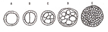
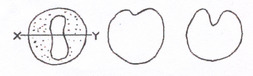
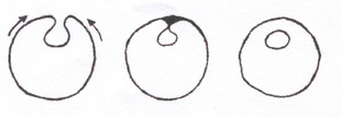
This is the only known congenital defect that occurs in the breed. (Congenital means that the defect is formed before birth). When considered as a defect in the dog family as a whole, Dermoid Sinuses occur only very rarely in dogs, other than Ridgebacks or Crossbred Ridgebacks. It must therefore be obvious that it is an inherited defect which has become widespread in the "blood lines" of the breed as a result of the early selective breeding of the original stock from which the Ridgebacks of today have been produced.The incidence of the defect throughout the breed is not known, as the recording of the numbers of Dermoid Sinus affected pups in litters has not been done on a scale large enough to enable a statistical analysis to be carried out. In fact, the occurrence of Dermoid Sinus affected pups on the litters of breeders has been kept confidential, as most breeders feel that there is considerable stigma attached to dogs and bitches amongst whose offspring Dermoid Sinus affected puppies occur.At this point I would like to state that with the present situation of breeding with selected outstanding dogs and bitches, no breeder without a program of progeny testing can be sure that his "blood lines" is free from the hereditary Dermoid Sinus. (The hereditary aspects of the condition will be dealt with in part two of this article). Thus, every purchased Ridgeback may be considered a potential carrier of the condition.THE FORMATION OF DERMOID SINUSTo understand the way, in which a Dermoid Sinus is formed, it is necessary to have some idea of how the embryo develops from a single fertilized egg cell in the womb of the bitch. Dermoid Sinus is a congenital defect that arises from a defect in the development of the embryo of a puppy.A fertilized egg resulting from a successful mating is a single simple cell. From this cell a puppy consisting of millions of specilized cells, which constitute the tissues and organs, must me formed in 63 days.
| ||||||||||||||